Etre comme on le désire tout simplement, grâce à la chirurgie & la médecine esthétique en Tunisie avec MEDHANNIBAL.
Double jaw surgery in Turkey
Double jaw surgery, medically known as Bi-Maxillary Osteotomy (or Bi-Max), is a complex orthognathic surgical procedure where both the upper jaw (maxilla) and the lower jaw (mandible) are surgically separated from the skull, repositioned, and fixed into a new alignment.
Unlike cosmetic plastic surgery which alters soft tissues, orthognathic surgery alters the skeletal foundation of the face. It is the intersection of dentistry, orthodontics, and maxillofacial surgery. While often associated with dramatic aesthetic changes, the primary goal is usually functional: correcting the bite (occlusion), opening the airway, and ensuring the jaws work in harmony.
This surgery is a major life event. It requires months (sometimes years) of preparation and a significant recovery period. This guide serves as a comprehensive resource for understanding the entire process.
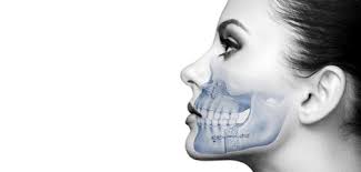
Anatomy and Physiology
To understand the surgery, one must understand the structures involved.
The Maxilla (Upper Jaw)
The maxilla forms the upper jaw and the central face. It houses the upper teeth, forms the floor of the nasal cavity, and the floor of the eye sockets (orbits). It is a stationary bone fused to the skull.
- Surgical Relevance: In double jaw surgery, the maxilla is cut horizontally (Le Fort I osteotomy). This allows it to be moved forward, backward, upward (impaction), or downward (downgrafting).
The Mandible (Lower Jaw)
The mandible is the only movable bone in the skull. It connects to the skull at the Temporomandibular Joints (TMJ).
- Surgical Relevance: The mandible houses the lower teeth and the Inferior Alveolar Nerve (IAN). The surgery involves splitting the bone to slide it forward or backward.
The Temporomandibular Joint (TMJ)
This is the hinge connecting the jaw to the temporal bones of the skull. It is one of the most complex joints in the body, allowing for rotation and translation (sliding).
- Surgical Relevance: Surgery alters the position of the mandible, which changes the pressure and seating of the condyles within the TMJ.
The Nerves
- Inferior Alveolar Nerve (IAN): Runs through the center of the lower jaw bone. It provides sensation to the lower lip and chin. It is the nerve most at risk during lower jaw surgery.
- Infraorbital Nerve: Located in the cheek area; provides sensation to the upper lip and nose.
Indications: Who Needs Surgery?
Double jaw surgery is prescribed when the skeletal discrepancy between the upper and lower jaws is too severe to be corrected by orthodontics (braces) alone.
Skeletal Malocclusions
- Class II Malocclusion (Retrognathism): The lower jaw is significantly set back compared to the upper jaw. This often results in a « weak chin » appearance and a deep overbite.
- Class III Malocclusion (Prognathism): The lower jaw protrudes beyond the upper jaw (underbite). This is common in certain genetic populations and creates a « concave » facial profile.
- Anterior Open Bite: The back teeth touch, but the front teeth do not overlap or touch. This makes biting into food (like a sandwich) impossible and often causes speech impediments (lisps).
- Asymmetry: One side of the jaw grows faster or longer than the other, causing the chin to deviate to one side and the dental midline to be off-center.
Functional Indications
- Obstructive Sleep Apnea (OSA): If the jaws are recessed, the airway is narrow. Moving both jaws forward opens the posterior airway space significantly.
- Masticatory Dysfunction: Inability to chew food properly, leading to digestive issues.
- TMJ Disorders: While surgery is not a guaranteed cure for TMJ pain, correcting the bite can alleviate stress on the joints in specific cases.
- Lip Incompetence: Inability to close the lips without straining the chin muscle (mentalis strain), often caused by vertical maxillary excess (long face syndrome).
The Pre-Surgical Phase
The journey usually begins 12 to 18 months before the actual surgery.
Step 1: Orthodontic Decompensation
This is a counter-intuitive phase. Over time, nature tries to camouflage a bad skeletal bite by tilting the teeth. For example, in an underbite, the lower teeth naturally tip backward to try to meet the upper teeth.
- The Process: The orthodontist uses braces to move the teeth into the correct position relative to the bone, not the opposing teeth.
- The Result: The bite and appearance often get worse during this phase. The underbite or overbite becomes more pronounced. This is necessary so the surgeon can move the bones maximally to fit perfectly.
Step 2: Medical Clearance
Patients undergo blood tests (CBC, clotting factors), chest X-rays, and EKG to ensure they can withstand general anesthesia.
Step 3: Virtual Surgical Planning (VSP)
Modern surgery rarely relies on 2D X-rays alone.
- CBCT Scan: A 3D Cone Beam CT scan captures the skull structure.
- Intraoral Scan: A digital wand scans the surface of the teeth.
- Simulation: Engineers and the surgeon merge these scans. They perform the surgery virtually on a computer, measuring movements down to the millimeter.
- Splint Fabrication: Based on the VSP, 3D-printed surgical splints (guides) are created. These acrylic distinct pieces fit over the teeth during surgery to lock the jaws into the exact planned position while plates are applied.
The Surgical Procedures (Technical Breakdown)
Double jaw surgery is performed under general anesthesia and typically takes 3 to 6 hours. All incisions are usually made inside the mouth (intraoral), leaving no external scars on the face (except for tiny stab incisions for screws in some cases).
A. The Upper Jaw: Le Fort I Osteotomy
- Incision: An incision is made inside the upper lip, high up in the gum line, extending from one side to the other.
- Exposure: The soft tissue is reflected to expose the maxilla.
- The Cut: A horizontal saw cut is made above the roots of the teeth and below the nose/sinuses.
- Down-fracture: The maxilla is carefully separated from the skull base.
- Mobilization: The maxilla is now loose. The surgeon can move it forward, up, or rotate it.
- Fixation: Once in the new position (guided by the surgical splint), the bone is secured using tiny titanium plates and screws. These usually stay in forever.
B. The Lower Jaw: Bilateral Sagittal Split Osteotomy (BSSO)
- Incision: Incisions are made behind the back molars on both sides.
- The Cut: The surgeon makes precise cuts in the ramus (the vertical part of the jaw). The cut is designed to split the bone lengthwise (like splitting a bagel).
- The Split: A chisel is used to separate the inner and outer segments of the bone.
- Crucial Step: The Inferior Alveolar Nerve runs through this bone. The surgeon must ensure the nerve remains intact in the inner segment while the outer segment (bearing the teeth) is moved.
- Movement: The tooth-bearing segment is slid forward or backward.
- Fixation: Titanium screws or plates are driven through the overlapping bone segments to hold them rigid.
C. The Chin: Genioplasty (Optional but Common)
Often, moving the jaws leaves the chin looking unbalanced. A genioplasty is a cosmetic addition to the functional surgery.
- The Cut: A cut is made below the lower front teeth roots.
- Movement: The chin button is moved forward, backward, or vertically.
- Fixation: Secured with a plate and screws.
The Hospital Experience
The Day of Surgery
- Anesthesia: You will be completely asleep. Because the surgery is in the mouth, the breathing tube (intubation) is placed through the nose (nasotracheal intubation).
- Catheter: A urinary catheter is usually placed as you will be immobile for hours.
Immediate Post-Op (The ICU/PACU)
Waking up is often the most difficult part.
- Congestion: Your nose will be blocked due to dried blood and swelling. Since your jaws are banded shut, this can cause panic. You must learn to breathe calmly through the gaps in your teeth or a small airway tube.
- Swelling: Swelling begins immediately.
- Numbness: Your entire lower face will likely be numb. You won’t feel your lips, chin, or tongue.
- Banding: Your jaws will be held together with tight rubber bands (elastics). Rigid wiring (wiring the jaw shut) is rare nowadays; elastics allow for emergency opening if necessary.
Hospital Stay
Most patients stay 1 to 3 nights.
- Pain Management: IV morphine or fentanyl is used initially, transitioning to liquid oral painkillers. Surprisingly, bone pain is not severe; the discomfort comes from swelling and muscle tension.
- Steroids: High-dose IV steroids are given to control swelling.
- Ice: Jaw bra (ice packs wrapped around the face) is applied constantly.
Post-Operative Recovery: The Timeline
Recovery is a marathon, not a sprint.
Week 1: The Survival Mode
- Swelling: Peaking around Day 3 or 4. You may look unrecognizable. The lips may become massive.
- Diet: Strictly liquid. Syringes or squeeze bottles are used because you cannot suck on a straw (suction creates negative pressure which can disrupt clots) and you cannot open your mouth.
- Hygiene: You cannot brush. You will use a prescription Chlorhexidine rinse to keep the mouth clean.
- Energy: Very low. Sleeping requires being propped up at a 45-degree angle to reduce swelling.
- Nose Bleeds: Minor oozing from the nose is common as the sinuses clear out (from the upper jaw surgery). Do not blow your nose.
Weeks 2-4: The Turning Point
- Swelling: Begins to subside. Bruising (yellow/green) appears on the neck and chest.
- Elastics: The surgeon may loosen the rubber bands, allowing you to open your mouth a few millimeters.
- Diet: Transition to « no-chew » puree. Anything that can be swallowed without chewing (mashed potatoes, blended soups).
- Nerves: You may start feeling « tingling » or « shocks » in the chin. This is a good sign—it means the nerves are waking up (regeneration).
Weeks 6-12: Bone Healing
- Bone Union: By week 6, the bones are usually fused enough to withstand mild force.
- Chewing: You are cleared to eat soft foods (scrambled eggs, soft pasta). No hard chewing (steaks, nuts) yet.
- Braces: Orthodontic adjustments resume to fine-tune the bite now that the bones are in place.
Months 6-12: Finalization
- Swelling: The final 10-20% of residual swelling (especially in the cheeks and nose tip) takes a full year to resolve.
- Hardware Removal: Generally, plates stay in. However, if they cause infection or irritation, they are removed in a minor outpatient procedure around month 9-12.
Diet and Nutrition Guide
Weight loss is almost guaranteed (usually 5-10% of body weight). Maintaining calorie intake is crucial for bone healing.
Liquid Phase (Week 1)
- Tools: 60ml catheter tip syringes, condiment squeeze bottles.
- Foods:
- High-calorie protein shakes (Ensure, Boost).
- Bone broth.
- Blended fruit juices (strained).
- Water (hydration is critical).
Puree Phase (Weeks 2-5)
- Rule: If you can squish it with your tongue against the roof of your mouth, you can eat it.
- Foods:
- Greek yogurt.
- Apple sauce.
- Mashed potatoes with gravy (add protein powder to gravy).
- Blended soups (clam chowder, tomato bisque).
- Scrambled eggs (very soft).
Soft Chew Phase (Week 6+)
- Foods:
- Fish (flaky).
- Overcooked pasta.
- Pancakes.
- Soft cooked vegetables.
Supplements: Calcium, Vitamin D, and Vitamin C are recommended to accelerate bone fusion.
Risks and Complications
While generally safe, this is major surgery with inherent risks.
1. Nerve Injury (Paresthesia/Dysesthesia)
This is the most common complication.
- Mechanism: The Inferior Alveolar Nerve is stretched or compressed during the BSSO split.
- Outcome: Most patients experience temporary numbness. However, 5-15% of patients may have permanent numbness in a small patch of the chin or lip. It does not affect movement (motor nerves are different), only sensation.
2. Relapse
The muscles and soft tissues have « memory. » They may try to pull the jaws back to their original position.
- Prevention: Rigid fixation (plates/screws) and proper orthodontics minimize this.
- Risk Factors: Large movements (e.g., moving the jaw 10mm+) have higher relapse rates.
3. Infection
Because the incisions are inside the mouth (a bacteria-rich environment), infection can occur at the plate sites.
- Treatment: Antibiotics. If persistent, the hardware (plates) may need to be removed.
4. TMJ Issues
Surgery can improve TMJ pain, but in rare cases, it can worsen it or create new clicking/popping due to the change in condyle position.
5. Deviated Septum / Nasal Changes
Moving the upper jaw affects the nose base.
- Alar Base Cinch: Surgeons often put a stitch in the nose muscles to prevent the nostrils from widening too much.
- Septum: Sometimes the septum buckles when the maxilla is moved up; the surgeon usually corrects this simultaneously.
Double Jaw Surgery for Sleep Apnea
This is a specific subset of the surgery known as Maxillomandibular Advancement (MMA).
The Mechanism
In Obstructive Sleep Apnea (OSA), the tongue and soft tissues collapse into the throat during sleep. CPAP machines force air past this blockage. MMA surgery physically enlarges the airway.
The Procedure
- Both jaws are moved forward significantly (often 10mm or more).
- This pulls the tongue muscles and soft palate forward, creating a massive expansion of the posterior airway space.
Success Rates
MMA is the most effective surgical treatment for OSA, with success rates often cited between 85-95%. It is considered a cure for many, allowing them to stop using CPAP machines entirely.
Aesthetic Considerations for MMA
Because the movements are large, the aesthetic change is significant. Surgeons must balance the need for airway expansion with maintaining a harmonious facial profile (avoiding a « chimp-like » appearance from over-advancement).
Double jaw surgery is a transformative procedure. It restores the ability to bite into an apple, to sleep without gasping for air, and to smile with confidence.
However, the cost of admission is high: a rigorous preparation phase, a physically demanding recovery, and a test of mental resilience. Success relies on a « Triad of Cooperation »:
- The Surgeon: Precise planning and execution.
- The Orthodontist: Perfect dental alignment.
- The Patient: Strict adherence to hygiene, diet, and recovery protocols.
If you are considering this surgery, consult with a board-certified Oral and Maxillofacial Surgeon. Ask to see « before and after » photos of their specific cases, understand the risks regarding nerve sensation, and prepare your support system for the recovery weeks. For those suffering from severe functional deficits, the journey, while difficult, is almost always reported as « worth it » in the end.


