Etre comme on le désire tout simplement, grâce à la chirurgie & la médecine esthétique en Tunisie avec MEDHANNIBAL.
Winged blepharoplasty In Turkey : Redefining the Lateral Gaze
Blepharoplasty is a surgical procedure aimed at correcting various age-related changes around the eyes, such as droopy eyelids (dermatochalasis), puffy bags under the eyes (steatoblepharon), and fine lines. It can be performed on the upper eyelids, lower eyelids, or both. The primary objectives are typically aesthetic, enhancing the patient’s appearance by creating a more alert and youthful look, but it can also be functional, improving vision by removing skin that obstructs the visual field.
Traditional upper blepharoplasty involves an incision made within the natural crease of the upper eyelid, allowing for the removal of excess skin, muscle, and sometimes fat. Similarly, lower blepharoplasty can be performed via an incision just below the lash line (transcutaneous) or inside the eyelid (transconjunctival) to address fat prolapse and skin laxity.
Winged blepharoplasty, also known as lateral extended blepharoplasty or crow’s feet blepharoplasty, is a specialized variation that extends the upper eyelid incision laterally beyond the orbital rim, often into the temporal region. This « wing » or « tail » allows the surgeon to address a broader area of concern, specifically the excess skin and wrinkles (rhytids) that accumulate in the lateral canthal and temporal regions, commonly known as « crow’s feet. »
This technique is particularly beneficial when there is significant lateral hooding, descent of the lateral brow, or prominent lateral canthal rhytids that would not be adequately corrected by a standard blepharoplasty incision confined to the eyelid crease. It aims to provide a more comprehensive and harmonious rejuvenation of the entire periorbital area, often integrating with the natural lines of expression to camouflage the resulting scar.
Anatomy of the Periorbital Region
A profound understanding of the intricate anatomy of the periorbital region is paramount for any surgeon performing blepharoplasty, especially the winged variant. Precision in dissection and tissue handling directly correlates with aesthetic outcomes and the avoidance of complications.
Skin
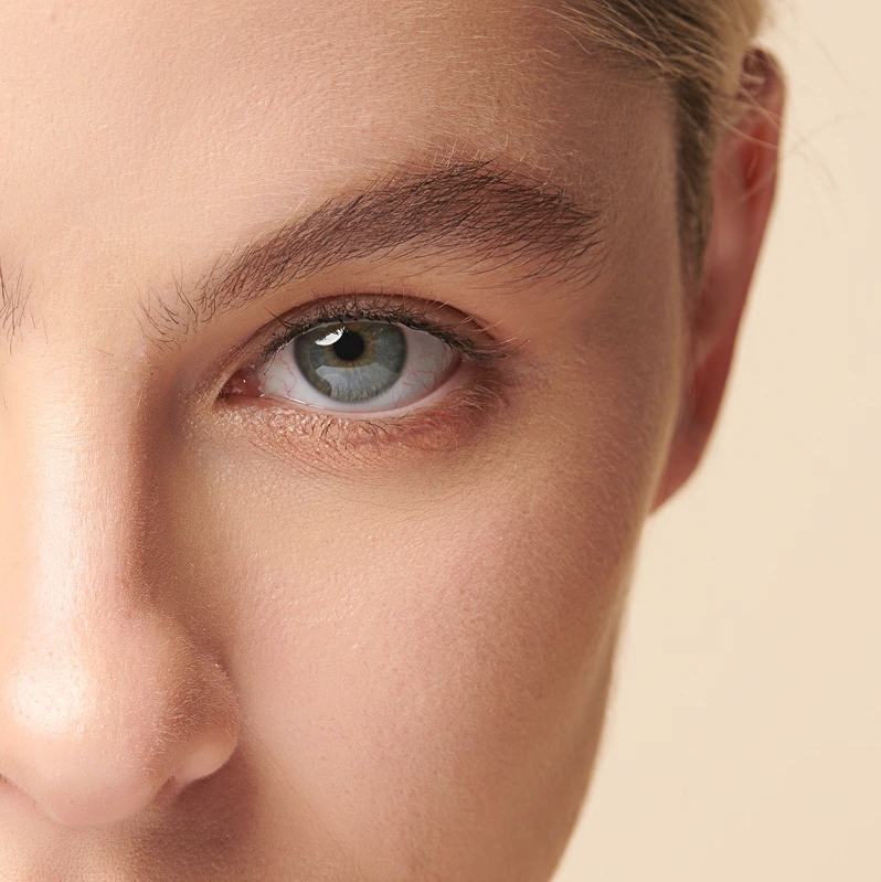
The skin around the eyelids is the thinnest on the entire body, making it particularly susceptible to the effects of aging, sun exposure, and repetitive muscle movements. Its delicate nature contributes to the early appearance of fine lines, wrinkles, and laxity (dermatochalasis). The lateral canthal area, where the « wing » incision is placed, is characterized by dynamic wrinkles (crow’s feet) that radiate outwards from the eye.
Muscles
The orbicularis oculi muscle is a critical component, responsible for eyelid closure and facial expressions. It is divided into three parts:
- Pretarsal part: Overlies the tarsal plate, responsible for involuntary blinking.
- Preseptal part: Overlies the orbital septum, responsible for voluntary blinking and gentle eye closure.
- Orbital part: Extends beyond the orbital rim into the temporal and cheek regions, responsible for forceful eye closure and contributing significantly to crow’s feet. In winged blepharoplasty, this orbital part is often targeted for modification or partial excision to address lateral rhytids.
Other muscles in the vicinity include the corrugator supercilii (responsible for vertical frown lines between the eyebrows) and the procerus (responsible for horizontal lines at the root of the nose), which are sometimes addressed in conjunction with blepharoplasty or brow lifts.
Orbital Septum
A thin, fibrous membrane that originates from the orbital rim and extends to the tarsal plates in both upper and lower eyelids. It acts as a barrier, holding the orbital fat within the orbit. Weakening or dehiscence of the septum allows orbital fat to prolapse forward, creating the characteristic « bags » or puffiness.
Orbital Fat Pads
The orbital fat is organized into distinct compartments:
- Upper Eyelid: Typically two fat pads – a medial (often paler, more fibrous) and a central (yellowish). A small, often overlooked, lateral fat pad may also be present, sometimes associated with the lacrimal gland.
- Lower Eyelid: Three fat pads – medial, central, and lateral. The medial fat pad is usually the most prominent.
Proper identification and conservative management (repositioning rather than aggressive excision) of these fat pads are crucial to avoid a hollowed-out appearance.
Tarsal Plate
Dense connective tissue that forms the structural support for the eyelids. The upper tarsus is larger and more rigid than the lower.
Levator Aponeurosis
The fibrous expansion of the levator palpebrae superioris muscle, responsible for elevating the upper eyelid. Damage to this structure can lead to ptosis (drooping of the upper eyelid).
Lacrimal Gland
Located in the superotemporal aspect of the orbit, responsible for tear production. It can sometimes prolapse, mimicking a lateral fat pad, and must be differentiated during surgery.
Canthal Tendons
- Medial Canthal Tendon (MCT): Anchors the eyelids medially to the nasal bone.
- Lateral Canthal Tendon (LCT): Anchors the eyelids laterally to the lateral orbital rim. Its integrity and position are crucial for maintaining proper eyelid tension and shape. Laxity or descent of the LCT contributes to a rounded or « sad » eye appearance and can be addressed during winged blepharoplasty via canthopexy or canthoplasty.
Nerves
Several nerves are at risk during periorbital surgery:
- Facial Nerve (CN VII): The temporal branch of the facial nerve is particularly vulnerable in the temporal region, especially during lateral extensions of incisions or dissection. Injury can lead to temporary or permanent paralysis of the frontalis muscle (brow lift) or orbicularis oculi (eyelid closure).
- Sensory Nerves: Supraorbital, supratrochlear, infratrochlear, zygomaticofacial, and infraorbital nerves provide sensation to the periorbital area. Injury can result in numbness or paresthesia.
Blood Vessels
The periorbital region is highly vascular. Key vessels include:
- Branches of the ophthalmic artery (supraorbital, supratrochlear, dorsal nasal).
- Angular artery (medial canthus).
- Superficial temporal artery (temporal region).
- Infraorbital artery.
Meticulous hemostasis is essential to prevent hematoma formation, which can be a sight-threatening complication (retrobulbar hematoma).
Bony Orbit
The bony confines of the orbit protect the globe and serve as attachment points for ligaments and muscles. The orbital rim defines the boundary between the eyelid and the surrounding facial structures.
Understanding the Aging Periorbital Region and Indications for Winged Blepharoplasty
The aging process manifests uniquely in the periorbital region, often necessitating a tailored surgical approach. Winged blepharoplasty is specifically indicated for a subset of patients presenting with particular patterns of aging.
General Signs of Periorbital Aging
- Dermatochalasis: Excess, lax skin on the upper eyelids, often creating a hooding effect that can sometimes impair peripheral vision.
- Steatoblepharon: Protrusion of orbital fat, leading to puffiness or « bags » in the upper and/or lower eyelids.
- Rhytids (Wrinkles):
- Crow’s Feet: Dynamic and static wrinkles radiating from the lateral canthus, caused by repetitive contractions of the orbital part of the orbicularis oculi muscle and skin laxity.
- Fine lines on the eyelids.
- Brow Ptosis: Drooping of the eyebrows, which can exacerbate upper eyelid hooding. It’s crucial to differentiate true dermatochalasis from pseudodermatochalasis caused by brow ptosis.
- Lateral Canthal Descent/Rounding: The outer corner of the eye may droop or lose its acute angle, contributing to a « sad » or tired appearance.
- Tear Trough Deformity: A hollow or groove extending from the inner corner of the eye obliquely across the lower eyelid, often due to volume loss and ligamentous attachments.
- Scleral Show: Excessive visibility of the white part of the eye below the iris, often associated with lower eyelid laxity or previous aggressive blepharoplasty.
- Volume Loss: Atrophy of fat and bone in the periorbital and temporal regions can lead to a hollowed, gaunt appearance.
Specific Indications for Winged Blepharoplasty
Winged blepharoplasty is particularly advantageous in cases where:
- Significant Lateral Hooding and Skin Excess: When the excess skin of the upper eyelid extends significantly beyond the lateral orbital rim and cannot be adequately addressed by an incision confined to the eyelid crease. This lateral skin often contributes to a heavy, tired look.
- Prominent Crow’s Feet (Lateral Canthal Rhytids): For patients with deep, static crow’s feet that are not fully amenable to non-surgical treatments (like neurotoxins) or require direct excision for a more dramatic improvement. The extended incision allows for direct removal of skin and sometimes a portion of the underlying orbicularis muscle in this area.
- Desire for Lateral Canthal Lift or Repositioning: The extended incision provides excellent access to the lateral canthal tendon, facilitating canthopexy (suture suspension) or canthoplasty (reconstruction) to elevate and reshape the lateral canthus, correcting a rounded or downward-sloping eye corner. This can help create a more almond-shaped eye.
- Concomitant Brow Ptosis (Lateral): While not a substitute for a formal brow lift, the lateral extension can provide a modest lift to the tail of the eyebrow by excising skin and suspending tissues in the temporal region. It can be a complementary procedure to a brow lift or sufficient for mild lateral brow descent.
- Correction of « Sad Eye » Appearance: By addressing lateral hooding and supporting the lateral canthus, the procedure can transform a tired or « sad » eye appearance into a more alert and youthful one.
- Revision Blepharoplasty: In cases where previous blepharoplasty failed to adequately address lateral skin excess or resulted in a rounded lateral canthus, the winged approach can be used to refine the results.
- Ethnic Considerations: In some ethnic groups, the natural eyelid crease may be lower or less defined, and the lateral extension can be adapted to create a desired shape or address specific concerns while respecting anatomical variations.
Preoperative Assessment and Planning
Meticulous preoperative assessment and planning are the cornerstones of a successful winged blepharoplasty, ensuring patient safety, realistic expectations, and optimal aesthetic and functional outcomes.
Patient Consultation and History
- Patient Expectations: A thorough discussion about the patient’s goals, concerns, and desired outcomes is crucial. It’s important to establish realistic expectations and explain the limitations of the procedure.
- Medical History:
- Ophthalmologic History: Dry eyes (xerophthalmia), glaucoma, detached retina, previous eye surgery, contact lens use. Patients with dry eyes may experience exacerbation post-surgery.
- Systemic Diseases: Thyroid disorders (Graves’ ophthalmopathy can cause eye bulging), hypertension, diabetes, bleeding disorders, autoimmune diseases.
- Medications: Aspirin, NSAIDs, anticoagulants, herbal supplements (e.g., ginkgo biloba, ginseng, vitamin E) can increase bleeding risk and should be discontinued well in advance.
- Smoking: Impairs healing and increases complication rates.
- Previous Facial Surgery: Prior blepharoplasty, brow lift, facelift, or laser resurfacing can affect tissue characteristics and scarring.
Ophthalmologic Evaluation
A comprehensive eye exam is essential to rule out pre-existing conditions that could be exacerbated by surgery or affect the outcome.
- Visual Acuity: Baseline measurement.
- Visual Fields: To document any pre-existing visual obstruction due to dermatochalasis.
- Schirmer’s Test: Measures tear production. Patients with low tear production are at higher risk for postoperative dry eye syndrome.
- Intraocular Pressure: To screen for glaucoma.
- Corneal Sensitivity: Important for patients with contact lenses or previous corneal issues.
Facial Analysis
A detailed aesthetic analysis of the entire face, not just the eyelids, is critical for harmonious results.
- Skin Quality and Elasticity: Assesses the skin’s ability to redrape and heal.
- Brow Position: Evaluate for brow ptosis. If significant, a brow lift may be recommended as a standalone procedure or in conjunction with blepharoplasty, as addressing only the eyelids will not correct a drooping brow. The lateral tail of the brow is particularly relevant for winged blepharoplasty.
- Upper Eyelid Crease: Note its height, definition, and symmetry.
- Lash Position: Assess for entropion (inward turning) or ectropion (outward turning).
- Amount of Skin Excess: Quantify the skin redundancy in the medial, central, and especially the lateral aspects of the upper eyelid and temporal region.
- Fat Prolapse: Identify the presence and location of orbital fat herniation.
- Lateral Canthal Position and Integrity: Assess for canthal dystopia (malposition), laxity (snap test, distraction test), and rounding. This is a key determinant for combining winged blepharoplasty with canthopexy/canthoplasty.
- Lower Eyelid Laxity: Perform a snap test (pulling the lower lid down and observing how quickly it returns to the globe) and a distraction test (pulling the lower lid away from the globe). Significant laxity often necessitates canthopexy/canthoplasty to prevent ectropion.
- Crow’s Feet: Evaluate their depth, extent, and whether they are static or dynamic.
- Symmetry: Note any pre-existing asymmetries, as perfect symmetry is rarely achievable.
Surgical Marking
This is a critical step performed with the patient in an upright, sitting position to accurately assess the effects of gravity and muscle tone.
- Upper Eyelid Crease: The primary incision line is marked within the natural upper eyelid crease, typically 8-10 mm above the lash line.
- Skin Excision Pattern: The amount of skin to be removed is carefully marked. The upper line of excision is determined by pinching the excess skin with forceps while ensuring complete eyelid closure.
- The « Wing » Extension: This is the defining feature of the procedure. The lateral extension of the incision is carefully planned to follow a natural crow’s foot line or a pre-existing wrinkle.
- It typically extends laterally and slightly upwards into the temporal region, often 1 to 2 cm beyond the orbital rim, sometimes up to 3-4 cm depending on the amount of skin to be excised.
- The exact shape and length of the « wing » are crucial for camouflaging the scar and achieving a natural lift. It should blend seamlessly with the temporal skin and avoid creating a noticeable « dog ear » deformity.
- The goal is to remove the lateral hooding and smooth out the crow’s feet while avoiding excessive tension.
- Fat Pads: Mark the areas of fat prolapse.
- Symmetry: Ensure markings are as symmetrical as possible.
Surgical Technique: Step-by-Step Winged Blepharoplasty
The execution of winged blepharoplasty requires meticulous surgical skill and an in-depth understanding of periorbital anatomy.
Anesthesia
Winged blepharoplasty can be performed under:
- Local Anesthesia with Sedation: This is a common approach, providing patient comfort while allowing for intraoperative patient cooperation (e.g., opening and closing eyes to check symmetry and closure).
- General Anesthesia: Preferred for longer procedures, combined procedures, or for anxious patients.
Local anesthetic (e.g., lidocaine with epinephrine) is infiltrated into the marked areas to provide analgesia and vasoconstriction, minimizing bleeding.
Incision Design (Upper Eyelid – Winged)
As marked preoperatively, the incision defines the boundaries of the skin to be excised.
- The lower incision line is made first, typically within the natural upper eyelid crease.
- The upper incision line is then made, meeting the lower line at the medial and lateral extents.
- The lateral « wing » is created by extending both the upper and lower incision lines outwards into the temporal region. The superior limb of the « wing » typically follows a natural crow’s foot line, while the inferior limb parallels it. The shape is often described as a « fishtail » or « exaggerated almond » shape. The precise angle and length of the wing are critical to achieve a natural-looking result and hide the scar effectively within existing rhytids.
Dissection and Tissue Management
- Skin Excision: The marked skin segment, including the lateral wing, is carefully excised using a scalpel or CO2 laser.
- Muscle Management (Orbicularis Oculi):
- Skin-Only Flap: In some cases, only skin is removed, leaving the underlying orbicularis muscle intact.
- Skin-Muscle Flap: More commonly, a strip of orbicularis muscle is removed along with the skin (skin-muscle flap). This can help reduce muscle bulk and further smooth out the lateral canthal area. In the lateral wing, a small portion of the orbital orbicularis muscle may be directly excised or plicated (tightened) to address crow’s feet and provide a modest lateral lift.
- Sub-orbicularis Dissection: For deeper rhytids, dissection beneath the orbicularis muscle can be performed to release attachments and allow for better redraping.
- Orbital Septum and Fat Management:
- The orbital septum is carefully incised to expose the underlying fat pads.
- Fat Excision: Prolapsed fat pads (medial and central in the upper eyelid) can be conservatively excised. Over-excision must be avoided to prevent a hollowed-out appearance.
- Fat Repositioning: In some modern techniques, fat is repositioned rather than excised, particularly in the lower eyelid, to fill hollows and create a smoother contour. In the upper eyelid, this is less common but can be considered.
- The lacrimal gland, if prolapsed, should be identified and repositioned, not excised.
Lateral Canthal Management (Often Combined)
The extended incision of winged blepharoplasty provides excellent access to the lateral canthal tendon, making it an ideal opportunity to address lateral canthal laxity or malposition.
- Lateral Canthopexy: This involves reinforcing the lateral canthal tendon by suturing it to the periosteum (fibrous membrane covering the bone) of the lateral orbital rim. This helps to elevate and support the lateral canthus, preventing rounding and contributing to a more almond-shaped eye.
- Lateral Canthoplasty: A more extensive procedure where the lateral canthal tendon is detached, shortened, and reattached to a higher position on the lateral orbital rim. This is used for more significant canthal malposition or laxity, or in revision cases.
Lower Eyelid Blepharoplasty (If Combined)
If lower eyelid concerns are also present, they are typically addressed at the same time.
- Transcutaneous Approach: An incision is made just below the lash line. A skin-muscle flap or skin-only flap is raised, and fat is managed (excised or repositioned). Excess skin is then trimmed. This approach provides direct access to skin and muscle.
- Transconjunctival Approach: An incision is made on the inside of the lower eyelid, avoiding an external skin incision. This is ideal for patients primarily concerned with fat bags and minimal skin laxity, as it leaves no visible scar. Fat can be excised or repositioned. If skin laxity is also an issue, a « skin pinch » excision (removing a small strip of skin below the lash line without undermining) can be combined with the transconjunctival approach.
- Orbicularis Suspension: In both lower lid approaches, the orbicularis muscle can be suspended laterally to provide additional support and lift to the lower eyelid.
Hemostasis
Throughout the procedure, meticulous hemostasis (control of bleeding) is paramount. Small blood vessels are coagulated using bipolar cautery. This prevents hematoma formation, which can be a serious complication.
Closure
- Layered Closure: Deeper tissues, such as the orbicularis muscle, may be closed with fine absorbable sutures to provide support and reduce tension on the skin.
- Skin Closure: The skin is closed with very fine, non-absorbable sutures (e.g., 6-0 or 7-0 nylon or prolene) in a continuous or interrupted fashion. The lateral « wing » incision requires particular attention to ensure a tension-free closure that aligns precisely with natural skin folds.
- Skin Adhesives/Tapes: Steri-Strips or tissue glue may be applied over the incisions for additional support and to optimize scar healing.
Postoperative Care
Proper postoperative care is crucial for minimizing complications, promoting healing, and achieving the best possible results.
- Immediate Post-Op:
- Cold Compresses: Applied to the eyelids for the first 24-48 hours to reduce swelling and bruising.
- Head Elevation: Sleeping with the head elevated helps reduce swelling.
- Pain Management: Mild pain relievers (e.g., acetaminophen) are usually sufficient. Avoid NSAIDs unless specifically cleared by the surgeon due to increased bleeding risk.
- Medications:
- Antibiotic Ointment/Drops: Applied to the incisions and/or eyes to prevent infection.
- Artificial Tears: To alleviate any temporary dry eye symptoms.
- Activity Restrictions:
- Avoid strenuous activities, heavy lifting, and bending over for at least 1-2 weeks to prevent increased blood pressure in the head and potential bleeding.
- Avoid rubbing the eyes.
- Avoid swimming and contact lenses until cleared by the surgeon.
- Suture Removal: Typically occurs 5-7 days after surgery.
- Expected Recovery:
- Swelling and Bruising: Common and usually peak within the first 2-3 days, gradually resolving over 2-4 weeks. The lateral « wing » area may experience more bruising due to the extended dissection.
- Discomfort: Mild to moderate discomfort is normal.
- Numbness/Altered Sensation: Temporary numbness around the incisions is common.
- Scarring: Initially red and slightly raised, scars typically fade over several months to a year, eventually becoming fine, white lines that are well-hidden within natural creases or crow’s feet lines.
- Follow-up: Regular follow-up appointments are scheduled to monitor healing and address any concerns.
Potential Complications
While winged blepharoplasty is generally safe, like any surgical procedure, it carries potential risks and complications. Patients must be fully informed of these possibilities.
General Surgical Risks
- Infection: Rare in eyelid surgery but possible.
- Bleeding/Hematoma: Collection of blood under the skin. Can be serious if it leads to a retrobulbar hematoma.
- Adverse Reaction to Anesthesia: Allergic reactions, respiratory issues.
Specific Complications of Blepharoplasty
- Ectropion (Lower Eyelid): Outward turning of the lower eyelid, exposing the conjunctiva. This is a significant concern, especially with lower blepharoplasty, and can lead to dry eyes, irritation, and cosmetic deformity. It is often prevented by meticulous skin excision, orbicularis suspension, and lateral canthal support.
- Lagophthalmos: Incomplete closure of the eyelids, leading to dry eyes and corneal exposure. Usually temporary but can be permanent if too much skin is removed.
- Dry Eyes: Exacerbation of pre-existing dry eye syndrome or new onset due to temporary changes in tear film or eyelid closure.
- Asymmetry: Minor asymmetries are common in natural anatomy and can persist or be created post-surgery. Significant asymmetry may require revision.
- Scarring: While scars are usually well-hidden, hypertrophic (raised) or keloid scars can occur, especially in individuals prone to them. The lateral « wing » scar, though designed to follow natural lines, can be more noticeable if not meticulously closed or if healing is suboptimal.
- Overcorrection/Undercorrection:
- Overcorrection: Too much skin/fat removed, leading to a hollowed-out or « stared » appearance, or lagophthalmos.
- Undercorrection: Insufficient tissue removal, leading to persistent hooding or bags.
- Ptosis (Eyelid Droop): Drooping of the upper eyelid, usually due to injury to the levator aponeurosis during surgery. Can be temporary or permanent.
- Vision Changes: Extremely rare but potentially devastating. A retrobulbar hematoma (bleeding behind the eyeball) can increase pressure on the optic nerve, leading to vision loss or blindness. This is a surgical emergency.
- Nerve Injury:
- Temporal Branch of Facial Nerve: Injury to this nerve during lateral dissection can result in temporary or permanent weakness of the frontalis muscle (brow movement) or the lateral orbicularis oculi (eyelid closure), leading to brow asymmetry or difficulty closing the eye.
- Sensory nerve injury can cause numbness.
- Persistent Crow’s Feet: While winged blepharoplasty aims to improve crow’s feet, some dynamic wrinkles will persist, and static ones may not be entirely eliminated. Adjuvant treatments like neurotoxins may still be beneficial.
- « Hollowed Out » Appearance: Can result from aggressive fat removal, especially in the lower eyelids.
- Pigmentation Changes: Discoloration of the skin, particularly in the lower eyelids.
Advantages and Disadvantages of Winged Blepharoplasty
Understanding the specific benefits and drawbacks helps in patient selection and managing expectations.
Advantages
- Comprehensive Lateral Rejuvenation: Effectively addresses excess skin and wrinkles (crow’s feet) that extend significantly beyond the lateral orbital rim, providing a more complete rejuvenation of the outer eye and temporal area than traditional blepharoplasty.
- Improved Eye Shape and Support: The extended incision offers excellent access for performing lateral canthopexy or canthoplasty, which can lift and reshape the lateral canthus, creating a more youthful, almond-shaped eye and preventing lower eyelid laxity.
- Direct Excision of Crow’s Feet: Allows for direct removal of skin and sometimes muscle contributing to deep, static crow’s feet, offering a more pronounced and potentially longer-lasting improvement than non-surgical methods alone.
- Modest Lateral Brow Lift: Can provide a subtle lift to the tail of the eyebrow, especially when combined with orbicularis suspension in the temporal region, complementing a full brow lift or addressing mild lateral brow ptosis.
- Harmonious Results: By addressing the lateral periorbital unit as a whole, it can achieve a more balanced and natural-looking rejuvenation that integrates seamlessly with the rest of the face.
- Potentially Longer-Lasting Results: The more extensive tissue management in the lateral aspect may lead to more durable outcomes in this region compared to standard blepharoplasty.
Disadvantages
- Longer Incision/Scar: The primary drawback is the extended incision, which results in a longer scar compared to traditional blepharoplasty. Although designed to be hidden within natural lines, it can be more noticeable if healing is poor or if the design is imprecise.
- Increased Dissection: Involves a more extensive dissection of tissues in the lateral and temporal regions, potentially leading to more swelling, bruising, and a slightly longer recovery period in these areas.
- Higher Risk of Specific Complications: Due to the extended dissection, there’s a slightly increased risk of complications such as temporal branch facial nerve injury (though rare with careful technique) or visible scarring in the temporal area.
- Requires Specialized Expertise: This technique demands a surgeon with specific expertise in periorbital anatomy and advanced blepharoplasty techniques to ensure proper incision placement, tissue management, and complication avoidance.
- Not Suitable for All Patients: Patients with minimal lateral skin excess or those who primarily need brow elevation may be better candidates for traditional blepharoplasty or a standalone brow lift.
- Potential for « Pulled » Look: If excessive skin is removed or if the closure is under too much tension, there is a risk of an unnatural, « pulled » or « stared » appearance, particularly at the lateral canthus.
Comparison with Other Periorbital Rejuvenation Techniques
Winged blepharoplasty is one tool in a broader armamentarium for periorbital rejuvenation. Its selection often depends on the specific patient concerns and the overall facial aging pattern.
- Traditional Blepharoplasty: Focuses on skin and fat removal within the confines of the eyelid. Less effective for significant lateral hooding, crow’s feet, or lateral canthal descent.
- Brow Lift (Endoscopic, Coronal, Direct): Primarily addresses brow ptosis and forehead wrinkles. While a brow lift can improve upper eyelid hooding by repositioning the brow, it does not directly remove excess eyelid skin or address crow’s feet as effectively as winged blepharoplasty. Often combined with blepharoplasty for comprehensive upper facial rejuvenation.
- Canthopexy/Canthoplasty (as standalone or adjunct): Specifically target the lateral canthal position and tension. While they can be performed as standalone procedures, their integration into winged blepharoplasty offers synergistic benefits by providing direct access and addressing surrounding skin laxity.
- Fat Grafting (Autologous Fat Transfer): Used to restore volume in hollowed areas (e.g., tear troughs, temporal hollowing, upper eyelid sulcus). Can be a valuable adjunct to blepharoplasty to create a softer, more youthful contour and avoid a hollowed-out appearance.
- Laser Resurfacing: Improves skin texture, reduces fine lines, and tightens skin. Can be used as an adjunct to blepharoplasty to further refine skin quality around the eyes and reduce superficial wrinkles.
- Neurotoxins (e.g., Botox): Temporarily relax muscles to reduce dynamic wrinkles, particularly effective for crow’s feet, glabellar lines, and forehead lines. Often used in conjunction with blepharoplasty to optimize results and maintain them.
- Dermal Fillers: Used to fill hollows (e.g., tear troughs, temporal hollowing) and add volume. Provide temporary improvement and are non-surgical.
- Chemical Peels: Improve skin texture and reduce superficial wrinkles, similar to laser resurfacing but with different mechanisms and downtime.
The choice of procedure or combination of procedures is highly individualized, based on a comprehensive assessment of the patient’s anatomy, aging patterns, and aesthetic goals.
Winged blepharoplasty represents a sophisticated and highly effective surgical approach for patients presenting with significant lateral periorbital aging, including pronounced lateral hooding, deep crow’s feet, and lateral canthal descent. By extending the traditional blepharoplasty incision into the temporal region, it allows for a more comprehensive excision of redundant skin and muscle, direct management of lateral canthal issues, and a more harmonious rejuvenation of the entire eye area.
Achieving optimal results with winged blepharoplasty hinges upon several critical factors: a thorough understanding of the complex periorbital anatomy, meticulous preoperative assessment and planning, precise surgical execution, and diligent postoperative care. While the procedure offers distinct advantages in addressing specific lateral aging concerns, it also carries potential risks, including a longer scar and the need for specialized surgical expertise.
Ultimately, winged blepharoplasty, when performed by an experienced and skilled surgeon, can significantly enhance a patient’s appearance, restoring a more youthful, alert, and refreshed look that is both natural and long-lasting. It stands as a testament to the continuous evolution of aesthetic surgery, offering tailored solutions to meet the diverse needs of individuals seeking periorbital rejuvenation.


